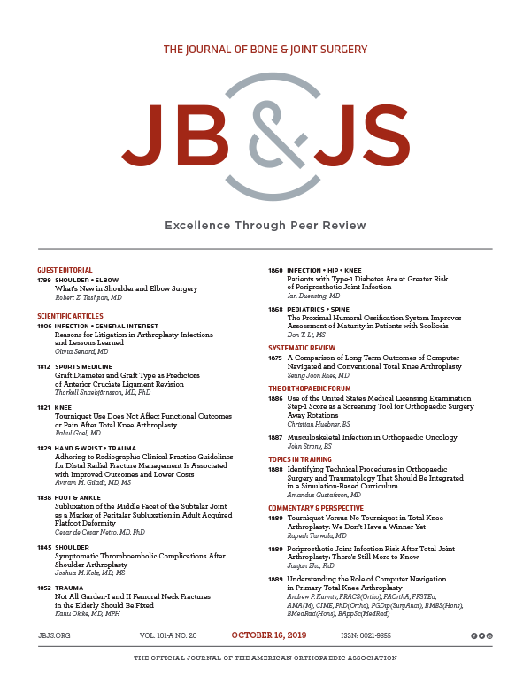
Patient-specific guides do not improve CT-assessed component alignment in TKA
Component alignment during total knee arthroplasty with use of standard or custom instrumentation: a randomized clinical trial using computed tomography for postoperative alignment measurement
J Bone Joint Surg Am. 2014 Mar 5;96(5):366-72Did you know you're eligible to earn 0.5 CME credits for reading this report? Click Here
Synopsis
63 male patients (64 knees) undergoing total knee arthroplasty (TKA) were randomized to receive treatment using either patient-specific cutting blocks - derived from 3D preoperative CT images - or standard instrumentation. The purpose of this study was to compare these two approaches with respect to component alignment and short-term clinical outcomes. Results at 6 months indicated that there were no significant differences between groups in regards to clinical outcomes or tibial and femoral component alignment. The number of outliers with respect to sagittal tibial alignment/slope was significantly greater when patient-specific guides were used.
Was the allocation sequence adequately generated?
Was allocation adequately concealed?
Blinding Treatment Providers: Was knowledge of the allocated interventions adequately prevented?
Blinding Outcome Assessors: Was knowledge of the allocated interventions adequately prevented?
Blinding Patients: Was knowledge of the allocated interventions adequately prevented?
Was loss to follow-up (missing outcome data) infrequent?
Are reports of the study free of suggestion of selective outcome reporting?
Were outcomes objective, patient-important and assessed in a manner to limit bias (ie. duplicate assessors, Independent assessors)?
Was the sample size sufficiently large to assure a balance of prognosis and sufficiently large number of outcome events?
Was investigator expertise/experience with both treatment and control techniques likely the same (ie.were criteria for surgeon participation/expertise provided)?
Yes = 1
Uncertain = 0.5
Not Relevant = 0
No = 0
The Reporting Criteria Assessment evaluates the transparency with which authors report the methodological and trial characteristics of the trial within the publication. The assessment is divided into five categories which are presented below.
1/4
Randomization
3/4
Outcome Measurements
3/4
Inclusion / Exclusion
4/4
Therapy Description
3/4
Statistics
Detsky AS, Naylor CD, O'Rourke K, McGeer AJ, L'Abbé KA. J Clin Epidemiol. 1992;45:255-65
The Fragility Index is a tool that aids in the interpretation of significant findings, providing a measure of strength for a result. The Fragility Index represents the number of consecutive events that need to be added to a dichotomous outcome to make the finding no longer significant. A small number represents a weaker finding and a large number represents a stronger finding.
Why was this study needed now?
A current trend in total knee arthroplasty (TKA) research is the use of patient-specific cutting blocks for improving the alignment of components. In order to customize these guides, patients undergo either magnetic resonance imaging (MRI) or computer tomography (CT) scans preoperatively to produce an image of their knee, from which these individualized cutting blocks are made. The majority of studies looking at this technique have used two-dimensional radiographs as opposed to 3D CT data. This study was needed to compare clinical outcomes and component alignment in patients undergoing TKA with either patient-specific cutting blocks (from 3D CT imaging) or standard instrumentation.
What was the principal research question?
In TKA, how does the use of patient-specific cutting blocks (derived from 3D preoperative CT images) compare to standard instrumentation (i.e. intramedullary femoral and external tibial cutting guides), with respect to clinical outcomes and component alignment, when assessed at 6 months?
What were the important findings?
- Between the study and control groups, respectively, there was no significant difference in surgical time (88.1 vs 92.1 minutes), postoperative hematocrit (31.9% vs 32.2%), hospital stay (3.1 vs 3.0 days), Knee Society rating scores (86.4 vs 90.2), Knee Society function scores (73.2 vs 82.1), improvement in KSS from baseline (+31.4 vs +31.1), or flexion arc (102.1 vs 104.1 degrees) (all p>0.05).
- Although one patient who underwent bilateral knee arthroplasty required two autologous units of blood, no patient in either group required a postoperative allogenic transfusion.
- There were no significant differences between the study and control groups with respect to the coronal mechanical axis (1.7 vs 1.3 degrees varus), the coronal femoral alignment (1.1 vs 1.0 degrees varus), femoral rotation (0.8 vs 1.7 degrees internal rotation), coronal tibial alignment (0.7 vs 0.3 degrees of varus), or sagittal tibial aslope (1.5 vs 2.4 degrees posterior) (all p>0.05).
- Although there were no significant differences between the study and control groups with respect to the percentage of outliers for the coronal mechanical axis (41% vs 38%), coronal femoral alignment (23% vs 23%), femoral rotation (27% vs 46%) and the coronal tibial alignment (14% vs 4%) (all p>0.05), there were significantly more outliers in the study group in regards to sagittal tibial alignment/slope (32% vs 8%; p=0.032).
- In the study group, the use of patient-specific guides was abandoned in 7/22 knees (32%). Insufficient extension space was noted in 12/22 knees (55%) of the study group, warranting additional cutting of either the femoral bone, the tibial bone, or both. Modifications to component size from preoperative plans in the study group occurred in 9/22 (41%) knees.
- In the control group, more bone was resected, following the initial cut, from either the distal femur or proximal tibia in 6 knees (23%) due to insufficient extension space. For one patient in this group, an excessive amount of bone was resected, requiring a polyethylene insert and a different, more constrained, implant.
- One patient in the control group required re-operation at 3 weeks, and another in the same group was scheduled to undergo revision for implant loosening at the time of publication.
What should I remember most?
In total knee arthroplasty, patient-specific guides were not associated with significant differences in surgical time, postoperative hematocrit, hospital stay, Knee Society Scores, range of motion, as well as tibial or femoral component alignment as compared with standard instrumentation. More cases where patient-specific guides were used had outliers in tibial slope.
How will this affect the care of my patients?
The results from this study suggest that the use of patient-specific cutting blocks from preoperative 3D CT scans do not improve femoral and tibial component alignment, and malalignment in tibial slope was more frequently observed with their use. This is an important finding since preoperative CT scans are costly and create a delay before surgery can be performed. As a result, further evaluation of efficacy is warranted, and future studies should include a cost-effectiveness analysis comparing patient-specific guides and conventional instrumentation.
Learn about our AI Driven
High Impact Search Feature
Our AI driven High Impact metric calculates the impact an article will have by considering both the publishing journal and the content of the article itself. Built using the latest advances in natural language processing, OE High Impact predicts an article’s future number of citations better than impact factor alone.
Continue
Join the Conversation
Please Login or Join to leave comments.