







SPORTS MEDICINE
A randomized controlled trial comparing bone mineral density changes of three different ACL reconstruction techniques
 This report has been verified
by one or more authors of the
original publication.
This report has been verified
by one or more authors of the
original publication.
OrthoEvidence Journal (OE Journal) - ACE Report
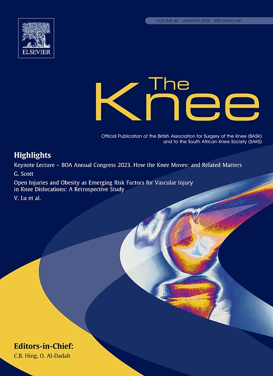
OE Journal. 2013;1(12):239 Knee. 2012 Dec;19(6):779-85. doi: 10.1016/j.knee.2012.02.005. Epub 2012 Mar 1662 male adult patients undergoing ACL reconstruction (ACLR) were randomized to either bone-patella tendon-bone (BPTB) graft, single-bundle hamstring (HT-SB) graft, or double-bundle hamstring (HT-DB) graft techniques. Changes in bone mineral density (BMD) of the three different ACLR techniques were measured at 1 day, 3 months, 5 months, and 1 year post-operation. Similar results were seen in the three ACLR techniques in terms of bone loss at the knee region, irreversible bone loss at the hip, and early clinical and functional outcomes up to 1 year after surgery. Additionally, a positive correlation existed between BMD at the distal femur and single-leg hop distance a 1 year post-operation.
Was the allocation sequence adequately generated?
Was allocation adequately concealed?
Blinding Treatment Providers: Was knowledge of the allocated interventions adequately prevented?
Blinding Outcome Assessors: Was knowledge of the allocated interventions adequately prevented?
Blinding Patients: Was knowledge of the allocated interventions adequately prevented?
Was loss to follow-up (missing outcome data) infrequent?
Are reports of the study free of suggestion of selective outcome reporting?
Were outcomes objective, patient-important and assessed in a manner to limit bias (ie. duplicate assessors, Independent assessors)?
Was the sample size sufficiently large to assure a balance of prognosis and sufficiently large number of outcome events?
Was investigator expertise/experience with both treatment and control techniques likely the same (ie.were criteria for surgeon participation/expertise provided)?
Oui = 1
Incertain = 0,5
Non pertinent = 0
Non = 0
L'évaluation des critères de rapport permet d'évaluer la transparence avec laquelle les auteurs rapportent les caractéristiques méthodologiques et les caractéristiques de l'essai dans la publication. L'évaluation est divisée en cinq catégories qui sont présentées ci-dessous.
3/4
Randomization
3/4
Outcome Measurements
4/4
Inclusion / Exclusion
4/4
Therapy Description
4/4
Statistics
Detsky AS, Naylor CD, O'Rourke K, McGeer AJ, L'Abbé KA. J Clin Epidemiol. 1992;45:255-65
L'indice de fragilité est un outil qui aide à l'interprétation des résultats significatifs, en fournissant une mesure de la force d'un résultat. L'indice de fragilité représente le nombre d'événements consécutifs qui doivent être ajoutés à un résultat dichotomique pour que le résultat ne soit plus significatif. Un petit nombre représente un résultat plus faible et un grand nombre un résultat plus fort.
Pourquoi cette étude était-elle nécessaire maintenant ?
Multiple studies have shown that after ACL reconstruction using BPTB graft, there can be a significant decrease in BMD around the knee region. However, many of these studies were neither randomized nor controlled, causing uncertainty in the reliability of the results. Additionally, none of these studies assessed BMD in HT-SB or HT-DB graft ACL reconstruction. This trial aimed to determine whether changes in BMD varied significantly between the ACL reconstruction techniques and elucidate the impact of BMD on the early functional or clinical outcomes.
Quelle était la principale question de recherche ?
Did BMD change around the knee region differ depending on the type of ACL reconstruction technique (BPTB, HT-SB, HS-DB) and did BMD loss at the knee region have a negative impact on the recovery of patients post-surgery when examined over a 1 year period?
Quels sont les résultats importants ?
- BMD at both the distal femur and proximal tibia decreased significantly at month 3 post-op (distal femur: -6.5% +/-6.9%; proximal tibia: -4.8% +/-7.0%) and month 5 post-op (distal femur: -7.3% +/-10.1%; proximal tibia: -5.6% +/-12.6%), increased and became insignificantly different from day 1 at 1 year post-op (distal femur: -1.6% +/-10.3%; proximal tibia: 0% +/-13.0%).
- There was no significant difference in BMD loss at the distal femur and proximal tibia among the three surgical techniques (p=0.205 and 0.121, respectively).
- There was significant irreversible loss of BMD at both the trochanteric region (month 3: -3.6% +/-3.3%; month 5: -4.5% +/-8.2%; 1 year: -4.2% +/-6.2%) and the femoral neck (month 3: -1.9% +/-3.7%; month 5: -2.4% +/-8.3%; 1 year: -2.5% +/-6.8%) of the injured limb.
- There was no significant difference in BMD loss among the three surgical techniques at the trochanteric region and femoral neck.
- At 1 year post-op, there were no significant differences between the different surgical techniques in IKDC score (p=0.759), Lysholm score (p=0.541), single-leg hop distance ratio (p=0.504), KT-1000 side-to-side difference (p=0.120), and manual Lachman test score (p=0.477)
- After combining data from the different groups, at 1 year post-op there was a significant improvement from baseline in IKDC score, Lysholm score, single-leg hop distance ratio, KT-1000 side-to-side difference, and manual Lachman test score (all p<0.001)
- Between BMD at the distal femur and the single-leg hop distance of the injured limb at 1 year post-op, a significant positive correlation was observed (r=0.299, p=0.031)
De quoi dois-je me souvenir en priorité ?
The results indicated that BMD loss was similar between BPTB graft, HT-SB graft, and HT-DB graft ACL reconstruction techniques. Additionally, early clinical and functional outcomes were similar among the three groups. As well, BMD at the distal femur and the single-leg hop distance a 1 year post-operation were positively related to one another.
Comment cela affectera-t-il les soins prodigués à mes patients ?
There appear to be no significant differences in BMD following these three types of ACL reconstruction in male patients. Further research is required to determine changes of BMD in female patients who have undergone different types of ACL reconstruction and if the type of rehabilitation has an effect. Moreover, the use of other high-resolution bone imaging systems, such as XtremeCT for in vivo BMD measurement can be incorporated in future studies in order to provide more data.

AVIS DE NON-RESPONSABILITÉ
Le contenu de cette page est fourni à titre d'information uniquement et n'est pas destiné à remplacer un avis médical, un diagnostic ou un traitement professionnel. Si vous avez besoin d'un traitement médical, demandez toujours l'avis de votre médecin ou rendez-vous au service des urgences le plus proche. Les opinions, croyances et points de vue exprimés par les individus sur le contenu de cette page ne reflètent pas les opinions, croyances et points de vue d'OrthoEvidence.
0 de 4 articles mensuels GRATUITS débloqués